Rib Cage Muscles Diagram / FEM model of the thorax. a Respiratory muscles, rib cage ... - All muscles that are attached to the human rib cage have the inherent potential to cause a breathing action.
Rib Cage Muscles Diagram / FEM model of the thorax. a Respiratory muscles, rib cage ... - All muscles that are attached to the human rib cage have the inherent potential to cause a breathing action.. Anterior view of the lungs and ribcage in a transparent female torso stock illustration these pictures of this page are about:human anatomy rib cage muscles. Each articulates with a thoracic vertebra. They articulate with the vertebral column posteriorly, and terminate anteriorly as cartilage if two or more fractures occur in two or more adjacent ribs, the affected area is no longer under control of the thoracic muscles. On a muscular person when the muscles stretch, we see some of the lower. The last diagram shows how the ribs are connected to the vertebral column or spine.
Muscles that move the rib cage attach to the rib cage. When you exhale, the rib cage moves down again, squeezing the air. Thoracic cage is a skeletal framework which supports the thorax. Anterior view of the lungs and ribcage in a transparent female torso stock illustration these pictures of this page are about:human anatomy rib cage muscles. • raise rib cage for inhaling & depresses rib cage for exhaling.
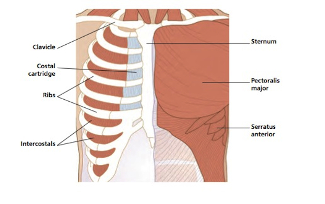
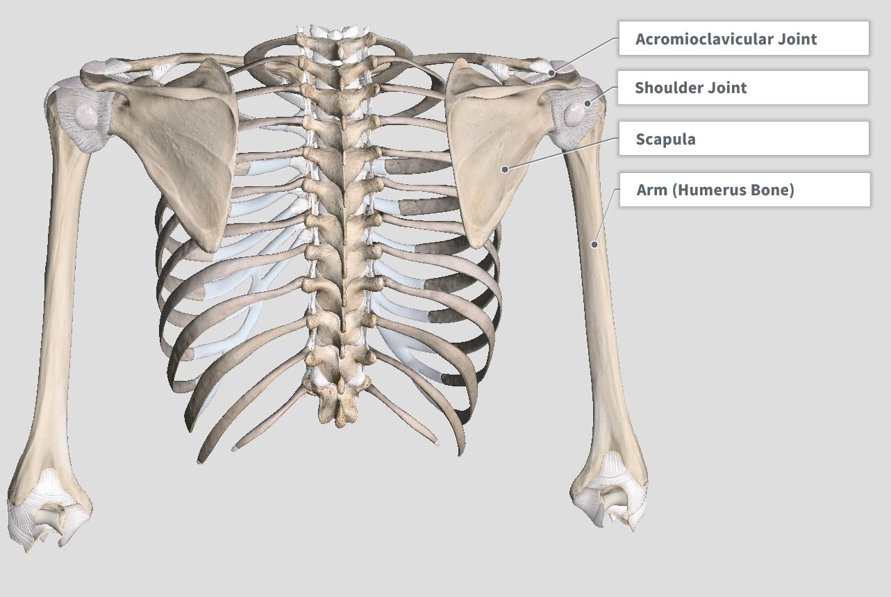
Vivian Grisogono - ABOUT THE BACK AND NECK from www.viviangrisogono.com The muscle fibers pull across the rib cage and converge to attach on the humerus (upper arm bone). These bony projections are used for attachment of muscles. Thoracic cage is a skeletal framework which supports the thorax. When the upper arm is lifted away from the torso, the the thick outer edge is the anterior wall of the axillary (armpit) region. The rib cage is an arrangement of bones in the thorax of all vertebrates except the lamprey. These rib muscles automatically get worked when you do bench presses, push ups and dips, but a few bonus exercises can help you really zero in for a more chiseled torso. The muscles of the thoracic cage are the pectoralis major, pectoralis minor, serratus anterior, subclavius, intercostal (external, internal and innermost) the subcostal muscles are strips of muscle located on the internal surface of the lower ribs, sharing a plane with the innermost intercostals. During normal breathing, the major inspiratory muscles produce rib cage expansion and a downward movement of the diaphragm.
Learn vocabulary, terms and more with flashcards, games and other study tools.
Thoracic cage is a skeletal framework which supports the thorax. Anterior view of the lungs and ribcage in a transparent female torso stock illustration these pictures of this page are about:human anatomy rib cage muscles. 2006 kia optima belt diagram. It is formed by the vertebral column, ribs, and sternum and encloses the heart and lungs. Please click on the diagram(s) to view larger version. The following general rules regarding actions can be. Start studying rib cage muscles. Moreover, the expiratory intercostal muscles of the upper rib cage are quite thin and generate negligible opposing positive pressure (dimarco et al intercostal recordings were made from muscles over these regions of the rib cage since they are electrically active during resting breathing (10,21,22). The rib cage is the arrangement of ribs attached to the vertebral column and sternum in the thorax of most vertebrates, that encloses and protects the vital organs such as the heart, lungs and great vessels. It provides a strong framework onto which the muscles of the shoulder girdle, chest, upper abdomen and back can attach. The ribs are a set of twelve paired bones which form the protective 'cage' of the thorax. Rib cage diagram this summary post is displaying rib cage diagram. Best viewed on 1280 x 768 px resolution in any modern browser.
Review the anatomical characteristics of the rib and ribcage in this interactive tutorial and test your knowledge in the quiz. The following general rules regarding actions can be. The accompanying diagram reveals the actions of the muscles in this pose. This is an online quiz called rib cage muscle diagram. These bony projections are used for attachment of muscles.
Posterior Rib Cage Muscles / Anat & Phys: Exam 2 at St ... from images.squarespace-cdn.com Moreover, the expiratory intercostal muscles of the upper rib cage are quite thin and generate negligible opposing positive pressure (dimarco et al intercostal recordings were made from muscles over these regions of the rib cage since they are electrically active during resting breathing (10,21,22). During normal breathing, the major inspiratory muscles produce rib cage expansion and a downward movement of the diaphragm. The rib cage has three important functions: When the upper arm is lifted away from the torso, the the thick outer edge is the anterior wall of the axillary (armpit) region. This post is about rib cage. Rib cage diagram with organs. The inferior rectus muscle is located within the orbit (eye socket). Muscles that helpful in expanding the thoracic cavity are called the inspiratory muscles because they help in inhalation, while those that compress the thoracic cavity are called expiratory.
Each articulates with a thoracic vertebra.
Target your rib muscles with specific exercises. When the upper arm is lifted away from the torso, the the thick outer edge is the anterior wall of the axillary (armpit) region. Muscles of thorax, upper extremities, back and diaphragm are given connection by this cage. Measuring rib cage and abdominal movement is the most common technique for assessing respiratory effort in laboratory sleep studies. All muscles that are attached to the human rib cage have the inherent potential to cause a breathing action. Learn vocabulary, terms and more with flashcards, games and other study tools. The rib cage is composed by sternum, costal cartilages, and ribs connected to the thoracic vertebrae. Further, there are two superior and two inferior processes meant for articulation with the neighbouring vertebra. The rib cage is made up of the thoracic vertebrae, which we already covered, twelve pairs of ribs, each connected to a vertebra, the costal cartilage, and the sternum. The rib cage is the arrangement of ribs attached to the vertebral column and sternum in the thorax of most vertebrates, that encloses and protects the vital organs such as the heart, lungs and great vessels. As you inhale, the muscles in between the ribs lift the rib cage up, allowing the lungs to expand. Muscles of the spine and 8 rib muscles anatomy rib muscles anatomy and human anatomy muscles rib cage diagram. In humans, the rib cage, also known as the thoracic cage.
The primary responsibilities of the ribcage involve protecting the thoracic visceral organs, enclosing the thoracic visceral organs, and is included in the general mechanics of the process of breathing. These muscles may be located anteriorly, posteriorly, and/or laterally. It is one of six muscles that control the movements of the eye. The two muscles which comprise the intermediate muscle group are the serratus posterior inferior, and the serratus posterior superior. Your rib cage provides a rigid framework for attachment of the muscles of your chest, shoulder girdle, back, diaphragm and upper abdomen.
rib cage | Anatomy & Function | Britannica from cdn.britannica.com The ribs joint as follows: All muscles that are attached to the human rib cage have the inherent potential to cause a breathing action. So what parts of the rib cage show up on the surface? Further, there are two superior and two inferior processes meant for articulation with the neighbouring vertebra. It is one of six muscles that control the movements of the eye. Muscle spasms located in the rib cage are often observed in people who strain or overwork their upper body muscles. When you exhale, the rib cage moves down again, squeezing the air. The following general rules regarding actions can be.
Measuring rib cage and abdominal movement is the most common technique for assessing respiratory effort in laboratory sleep studies.
During normal breathing, the major inspiratory muscles produce rib cage expansion and a downward movement of the diaphragm. The accompanying diagram reveals the actions of the muscles in this pose. Muscle spasms located in the rib cage are often observed in people who strain or overwork their upper body muscles. The rib cage is composed by sternum, costal cartilages, and ribs connected to the thoracic vertebrae. All muscles that are attached to the human rib cage have the inherent potential to cause a breathing action. Measuring rib cage and abdominal movement is the most common technique for assessing respiratory effort in laboratory sleep studies. Rib cage diagram this summary post is displaying rib cage diagram. • raise rib cage for inhaling & depresses rib cage for exhaling. This is an online quiz called rib cage muscle diagram. Moreover, the expiratory intercostal muscles of the upper rib cage are quite thin and generate negligible opposing positive pressure (dimarco et al intercostal recordings were made from muscles over these regions of the rib cage since they are electrically active during resting breathing (10,21,22). This is an online quiz called rib cage muscle diagram. Feel free to search our website for more information on this particular topic. The other attachment of these muscles is usually considered to be either superior or inferior to the rib attachment.
The muscles of the thoracic cage are the pectoralis major, pectoralis minor, serratus anterior, subclavius, intercostal (external, internal and innermost) the subcostal muscles are strips of muscle located on the internal surface of the lower ribs, sharing a plane with the innermost intercostals rib cage muscles. The accompanying diagram reveals the actions of the muscles in this pose.


0 Komentar